
Hematology Case Study - Leveraging RET-He to Identify Early Iron Deficiency
Download PDF
Latent iron deficiency, also termed iron-deficient erythropoiesis, represents a pre-anemic state in which iron stores are depleted but hemoglobin remains within reference range. Early identification is clinically significant, as the condition can rapidly progress to frank iron-deficiency anemia if affected individuals are not treated with iron supplementation. A sensitive, readily available hematology parameter for detecting this stage would therefore be beneficial. The reticulocyte hemoglobin equivalent (RET-He) has emerged as a reliable marker for assessing real- time iron availability for erythropoiesis, supporting both early detection and longitudinal monitoring.
Case Study*
A pregnant woman expecting twins presented to her physician for a scheduled evaluation. The routine full
blood count showed HGB, MCH and MCV values all within normal limits. In addition, the physician ordered a
reticulocyte panel. Results showed the RET-He was extremely low at 20.6 pg (1.28 fmol) suggesting evidence of
iron-deficient erythropoiesis. A low ferritin of 6.8 ng/mL subsequently confirmed that the patient had absolute
iron deficiency. Interestingly, the patient’s blood smear showed an overall normal morphology with very few
microcytic and hypochromic cells but no clear indication of iron deficiency.
In this case, routine CBC and peripheral blood smear morphology alone were insufficient to flag iron deficiency. Had RET-He not been included in the reticulocyte panel, iron-deficient erythropoiesis would likely have gone undetected and untreated. Given the physiologic demands of a twin pregnancy, unaddressed iron depletion carried significant risk — not only for maternal outcomes but for fetal development as well.
What is the Reticulocyte Hemoglobin Equivalent, or RET-He?
- RET-He is a reportable hematology parameter which reflects the hemoglobin content of reticulocytes (immature red blood cells).
- RET-He provides an early assessment of the available iron utilized in red blood cell production over the previous 2–4 days. 1,2
- RET-He reference range: 29.3–35.4 pg or 1.82–2.20 fmol. 3
- RET-He has been reported to have high accuracy, sensitivity and specificity for identifying iron deficiency. 4,5
- RET-He is readily available from a routine laboratory analysis of an EDTA blood sample.
*Results of case studies are not predictive of other cases and results may vary. It is the healthcare provider’s responsibility to determine applicability in routine clinical practice. Sysmex RET-He has been FDA cleared to classify and measure hemoglobin content of reticulocytes. The clinical applications or uses presented in these materials have not been cleared by the FDA.
Benefits of RET-He in Latent Iron Deficiency
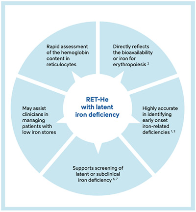
- Assesses the content of hemoglobin in reticulocytes.
- Reflects the bioavailability of iron for erythropoiesis. 1,2
- Supports detection of subclinical iron deficiency – the lack of iron before the onset of anemia. 6,7
References
[1] Hoenemann C et al. (2021): Reticulocyte and Erythrocyte Hemoglobin Parameters for Iron Deficiency and Anemia Diagnostics in Patient Blood Management. A Narrative Review. J Clin Med; 10(18): 4250.
[2] Hoenemann C et al. (2021): Reticulocyte Hemoglobin as a Routine Parameter in Preoperative Iron Deficiency Assessment. Endocrinol Metab; Vol. 5 No.1: 154.
[3] L van Pelt J et al. (2022): Reference intervals for Sysmex XN hematological parameters as assessed in the Dutch Lifelines cohort. Clin Chem Lab Med; 60(6): 907–920.
[4] Buttarello M et al. (2016): Evaluation of the hypochromic erythrocyte and reticulocyte hemoglobin content provided by the Sysmex XE-5000 analyzer in diagnosis of iron deficiency erythropoiesis. Clin Chem Lab Med; 54(12): 1939– 1945.
[5] Mehta S et al. (2016): Reticulocyte Hemoglobin vis- a-vis Serum Ferritin as a Marker of Bone Marrow Iron Store in Iron Deficiency Anemia. J Assoc Physicians India; 64(11): 38–42.
[6] Toki Y et al. (2017): Reticulocyte hemoglobin equivalent as a potential marker for diagnosis of iron deficiency. Int J Hematol;106(1): 116–125.
[7] Tiwari AK et al. (2018): Applying newer parameter Ret-He (reticulocyte hemoglobin equivalent) to assess latent iron deficiency (LID) in blood donors- study at a tertiary care hospital in India. Vox Sang; 113(7): 639–646.
Sigue leyendo Download PDF