Hematology Case Study - Micromegakaryocytes
Download PDF
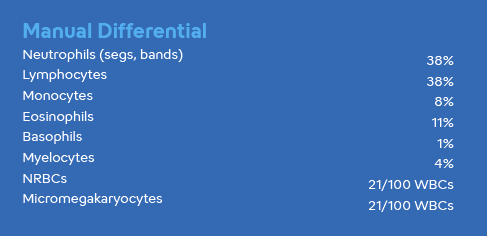
This newborn (see PDF) presented with leukocytopenia and thrombocytopenia with normal red cell indices. A high percentage of nucleated red cells were confirmed by manual smear review. An abnormal impedance platelet histogram suggested interference due to cell fragments or debris, resulting in an unreportable Mean Platelet Volume (MPV). However, a fluorescent PLT count (PLT-F) was performed, masking the “PLT Abn Distribution” flag. The PLT-F channel showed an increased Immature Platelet Fraction (IPF), indicating the presence of immature platelets in the peripheral blood.
Depending on the morphology of the micromegakaryocytes, these cells may or may not interfere with the WBC count. It is suggested that a WBC estimate and correction of the total nucleated cell count (using the TNC-N result found in the Service tab of the XN-Series™ Automated Hematology Analyzer) be performed, if necessary, according to your laboratory protocol. Please refer to the “NRBC Present” Message section of the XN-Series Flagging Interpretation Guide for further instruction.


The clinical applications or uses presented in these materials, including case studies, are provided for illustration purposes only. Prior to using any Sysmex device, please review the manufacturer’s instructions use. It is the healthcare provider’s responsibility to determine applicability in routine clinical practice.
Keep reading: Download PDF