Science of Urinalysis - Hemosiderin
Download PDF
What is Hemosiderin?
Hemosiderin is a granular form of hemoglobin that can be seen in the urine of patients with intravascular hemolysis. In conditions that cause hemolysis, such as paroxysmal nocturnal hemoglobinuria (PNH), hemoglobin is released from disrupted RBCs in the peripheral blood, then binds to haptoglobin for transport to the liver, where it will be cleared from the body. If the amount of extracellular hemoglobin exceeds the capacity of haptoglobin, the excess is released into the urine where it is partially reabsorbed in the renal tubules. In these tubular cells, the hemoglobin is converted to ferritin and then to hemosiderin, which is eventually released into the urine as the renal tubular cells degenerate.
Unstained hemosiderin is seen in the urine as yellowish-brown granules and may appear in macrophages, hemosiderin casts or be free-floating. Testing for urine hemosiderin usually involves concentrating the sample and staining the sediment with a Prussian blue solution, in which the hemosiderin will appear blue.
Urine hemosiderin testing may be performed to monitor patients with severe acute hemolytic anemia, PNH, hemochromatosis or in the case of an incompatible blood transfusion.
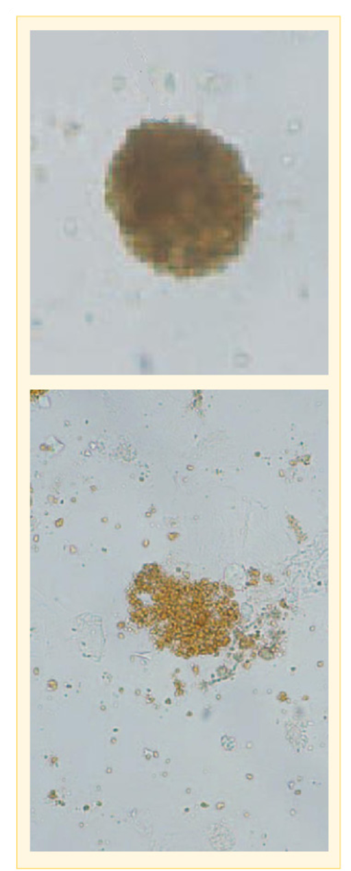
Morphology/Features:
- Small yellowish-red granules in unstained urine
- May be intracellular, free-floating or in hemosiderin casts
- Can resemble amorphous material
- Confirm with Prussian blue reaction
Resources:
Atlas of Urinary Sediment, Sysmex Corporation, Kobe, Japan.
Sigue leyendo Download PDF