
Hematology Case Study - Fragile WBCs
Download PDF
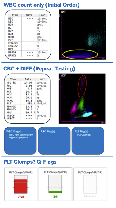
The patient is a 54-year-old female with metastatic breast cancer and new liver and bone metastasis who is currently undergoing chemotherapy. The original order on this patient was for a WBC only and no results were obtained on the XN-SeriesTM analyzer (shown at upper right). Repeat testing initiated by the lab showed a WBC count of 17.03 x 10³/µL (shown at center right).
In this sample, the extensive debris in the WNR channel (circled in yellow) caused the WBC result to be unreliable, so the result was masked (----). If a differential is part of the order, the XN can acquire the WBC count from the WDF channel, which is indicated by “&D” next to the WBC parameter. Note that the “WBC Abn Scattergram” flag can be generated by either of these scattergrams. Additionally, a “Difference between WNR and WDF” action message may be generated if the total nucleated cell count from each of these channels do not match within the acceptable limits. The counts obtained in the individual scattergrams – WBC-N (for WNR scatter) and WBC-D (for WDF scatter) – can be found on the “Service” tab in Data Browser.
Additional flagging on this sample includes the “PLT Clumps?” suspect flag. This flag can be generated in the WNR, WDF and PL T-F (not analyzed on this sample) channels. Some of the debris in the WNR scattergram is being classified as platelet clumps as demonstrated by the Q-Flags (shown at lower right).
NOTE: The individual Q-Flags for each of the channels can be added to the customizable Lab Only tab in Data Browser. See Chapter 11 of the XN Instructions for Use manual for instructions.
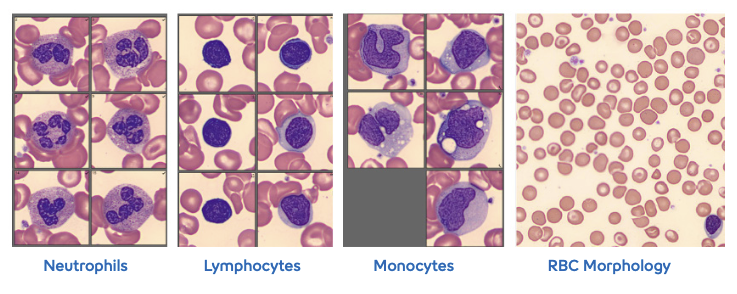
Review of the peripheral blood smear on the DI-60™ Automated Ditgital Cell Morphology Analyzer shows typical-looking neutrophils with no overt dysplasia, small round lymphocytes (some with scant cytoplasm) and reactive-appearing monocytes. Red cell morphology is remarkable for the presence of target cells and schistocytes. The WBC count obtained from the WDF channel can be verified. In addition, the XN detected a small population of WBCs with high fluorescence and moderate complexity (circled in red on the WDF scattergram). This population may represent the large, reactive monocytes shown below.
Although RBC fragments are present in the sample, this is not likely the source of the interference in the WNR scattergram. The lysing reagent used in the WNR channel is harsher than the reagent used in the WDF channel and can possibly cause WBC fragmentation; this may be why there is debris interference in the WNR channel but not in the WDF channel. The source of the fragile cells in this sample is likely due to chemotherapy treatment or circulating tumor cells.
Potential next steps for this specimen may include:
- Retest with WNR & WDF modes (CBC + DIFF) if not already ordered
- Perform WBC estimate by peripheral smear review
- Consult with lab pathologist to review clinical history, treatment, results and additional result comments (if needed)
The clinical applications or uses presented in these materials, including case studies, are provided for illustration purposes only. Prior to using any Sysmex device, please review the manufacturer’s instructions use. It is the healthcare provider’s responsibility to determine applicability in routine clinical practice.
Keep reading: Download PDF